
Skin Health Checks/ Skin Lesions

What is a skin health check and why does it matter?
- Full body dermoscopy examination ]
- Benign, precancerous and cancerous lesions
- Skin cancer detection and prevention
- Personalised risk based screening
A skin health check is a comprehensive, Consultant Dermatologist led examination of the entire skin surface, performed with dermoscopy, to identify and characterise any benign, precancerous, or cancerous skin lesions. The skin is the body's largest organ, and expert dermoscopic assessment is one of the most effective tools for the early detection of skin cancer.
During a skin health check, Dr Ophelia assesses for benign lesions including skin tags, benign moles (naevi), seborrhoeic keratoses, and dermatosis papulosa nigra, a benign condition causing small, raised dark spots that occurs predominantly in darker skin types. She also identifies precancerous changes, most commonly actinic keratoses, rough scaly lesions from cumulative sun damage that can progress to skin cancer if untreated, and solar lentigines, commonly known as sunspots or age spots. Most significantly, a skin health check is designed to detect skin cancer at the earliest possible stage, including non melanoma skin cancers such as basal cell carcinoma (BCC) and squamous cell carcinoma (SCC), and melanoma, the most serious form, where early diagnosis has the single greatest impact on outcome and survival.
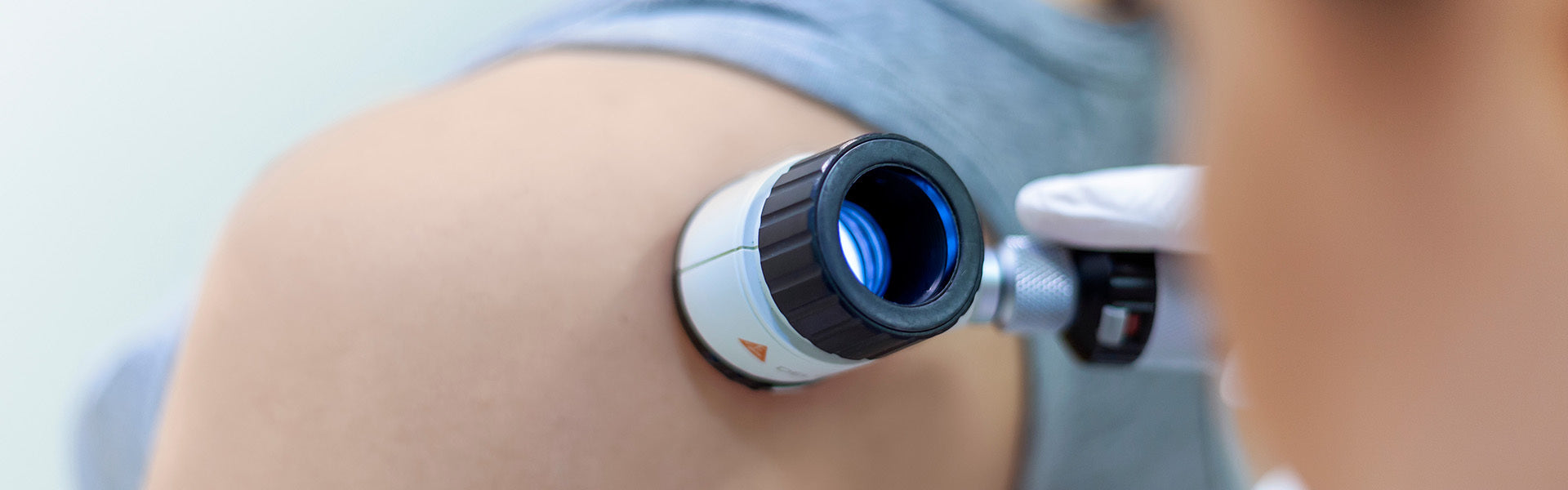
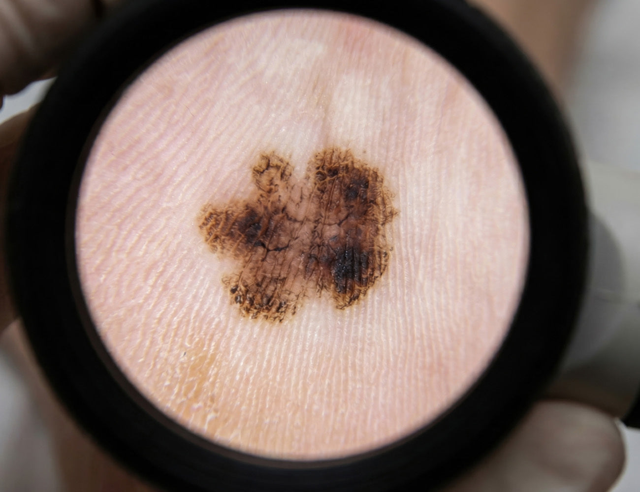
Dermoscopy, the use of a specialist handheld magnification device with polarised light, is central to a thorough examination, allowing visualisation of pigment patterns, vascular structures, and other features not visible to the naked eye. This improves accuracy in distinguishing benign lesions from precancerous or malignant ones and reduces unnecessary biopsies. Regular checks are recommended for anyone with risk factors including fair skin, a personal or family history of skin cancer, a high mole count, significant sun exposure or sunburn history, or a weakened immune system, though any new, changing, or concerning lesion warrants prompt specialist assessment.
What are my treatment options for skin lesions?
- Shave removal and punch biopsy
- Excision and wide local excision
- Electrocautery removal of benign skin lesions
- Topical chemotherapy creams
- IPL, resurfacing lasers (Clear+Brilliant and Fraxel) and photodynamic therapy
Treatment is tailored to the type, depth, and behaviour of each lesion, with the full range of procedural and medical options available within a single specialist practice.
For benign lesions such as skin tags, seborrhoeic keratoses, and dermatosis papulosa nigra, removal is most commonly achieved through shave removal under local anaesthetic, or electrocautery, which uses controlled heat to destroy superficial growths. Both are quick, well tolerated, and produce excellent cosmetic results, with particular care in darker skin types to manage the risk of post inflammatory pigment change.
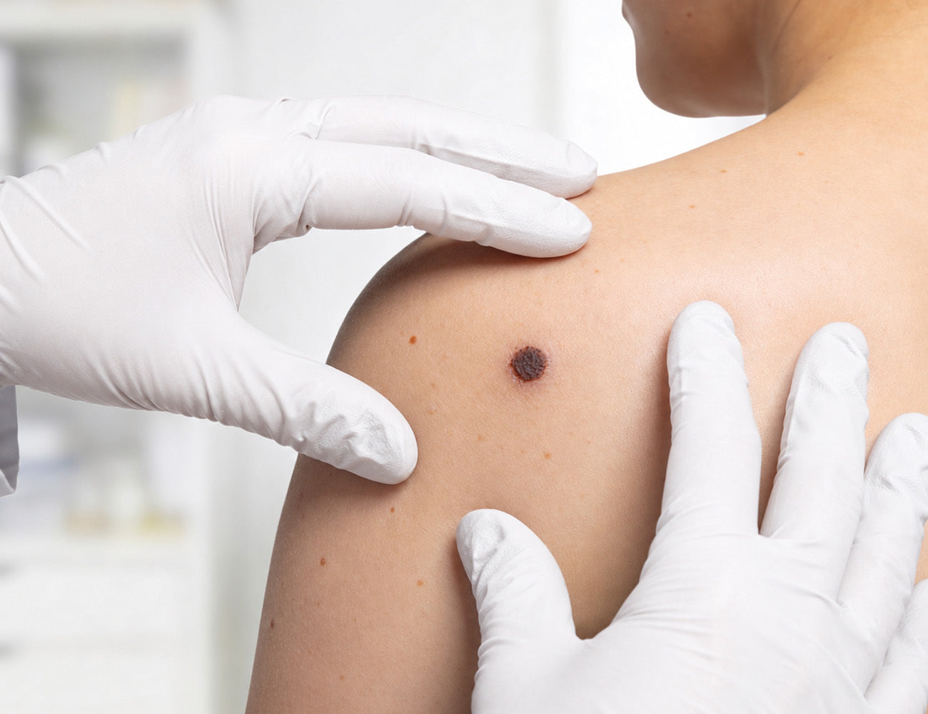
Where a lesion is suspicious or a histological diagnosis is required, Dr Ophelia performs diagnostic punch biopsies for laboratory analysis under local anaesthetic. For lesions confirmed or strongly suspected to be skin cancer, surgical excision removes the lesion with an appropriate margin of normal skin, and for more advanced or higher risk skin cancers, a wide local excision ensures complete clearance and minimises recurrence. Having a Consultant Dermatologist perform the full pathway, from dermoscopic assessment through biopsy to definitive surgery, in one coordinated setting reduces delays and ensures continuity of care from diagnosis to cure.
For precancerous changes including actinic keratoses, effective medical and procedural treatments vary with the extent of the affected skin. Prescription topical agents, including diclofenac gel (Solaraze), imiquimod, and 5-fluorouracil (Efudix), selectively target abnormal, sun-damaged cells across a treatment area, making them particularly valuable for field cancerisation, where widespread subclinical UV damage affects a broad area of skin. Laser and light based treatments, including IPL, Picosure Pro, Clear + Brilliant, and Fraxel Dual resurfacing laser, are also highly effective for actinic keratoses, solar lentigines, and sun damage, while improving skin texture and tone. For extensive or treatment resistant cases, photodynamic therapy (PDT) uses a photosensitising agent activated by light to selectively destroy abnormal cells.
Why should I see Dr Ophelia Veraitch for a skin health check and skin lesion treatment in London?
- Award winning consultant dermatologist
- Full body dermoscopic examination and diagnosis
- Full procedural range. Biopsy, excision, wide local excision, electrocautery
- Prescription field treatment; Solaraze, imiquimod, Efudix
- PDT for AKs, IPL, Clear + Brilliant, Fraxel Dual in house laser treatment for sun damage
As an award winning Consultant Dermatologist, Dr Ophelia Veraitch performs every skin health check personally, using careful dermoscopic examination to assess benign lesions, identify precancerous change, and detect skin cancer at the earliest stage.
Her expertise covers the complete pathway, from shave removals and electrocautery for benign lesions, with particular care in darker skin types, to punch biopsies and surgical excision or wide local excision where skin cancer is confirmed. Having this full pathway under one specialist means patients move from initial concern to definitive management with minimum delay.
Dr Ophelia understands that any new or changing skin lesion can cause worry, and she communicates findings clearly, explains the reasoning behind any recommended biopsy or treatment, and ensures every patient leaves with a clear plan for follow up.
HELP
Frequently Asked Questions - Skin Health Checks And Skin Lesions
Frequency depends on risk factors; higher-risk patients should be checked at least annually and those with a previous skin cancer more frequently, while any new, changing, or symptomatic lesion warrants prompt assessment.
Dermoscopy uses a specialist handheld magnification device with polarised light to reveal pigment patterns, vascular structures, and features invisible to the naked eye, improving accuracy in distinguishing benign from precancerous or malignant lesions.
Dermatosis papulosa nigra is a common benign condition causing small, raised, dark spots, predominantly in darker skin types, treated with shave removal or electrocautery with particular care to minimise post inflammatory hyperpigmentation.
An actinic keratosis (AK) is a rough, scaly, precancerous lesion from cumulative UV damage that can progress to squamous cell carcinoma if untreated, and often signals wider subclinical sun damage, or field cancerisation.
A punch biopsy removes a small cylindrical sample for analysis; an excision removes a lesion with a margin of normal skin; and a wide local excision takes a larger margin for more advanced or higher-risk skin cancers, including melanoma.
Electrocautery uses controlled heat through a fine probe to destroy superficial benign lesions such as skin tags, seborrhoeic keratoses, and dermatosis papulosa nigra, but is not used for suspicious lesions as it does not preserve tissue for histology.
Field cancerisation is a large area of sun-exposed skin with widespread subclinical UV damage of which actinic keratoses are the visible tip; it is treated across the whole field with prescription topical agents including 5-fluorouracil (Efudix), imiquimod, or diclofenac gel (Solaraze), or photodynamic therapy (PDT).
Photodynamic therapy applies a photosensitising cream absorbed into abnormal cells, which is then activated by light to selectively destroy precancerous cells. It is particularly effective for widespread or treatment-resistant actinic keratoses and field cancerisation.
Laser and light-based treatments including IPL, Clear + Brilliant, and Fraxel Dual address sun damage, and emerging evidence suggests fractional resurfacing may reduce subsequent actinic keratoses, though it is not a substitute for regular checks and sun protection.
Any mole that is new in adulthood, changing in size, shape, or colour, developing irregular borders, becoming raised, bleeding, or itching should be assessed promptly; the ABCDE rule, Asymmetry, Border irregularity, Colour variation, Diameter greater than 6mm, and Evolution, guides initial concern, but early assessment and biopsy is the single most important factor in outcome.
A Consultant Dermatologist has specialist training in dermoscopy and the diagnosis of benign, precancerous, and malignant lesions, plus the surgical skills for biopsies and excisions and the authority to prescribe topical chemotherapy and photodynamic therapy, which GPs and non medical skin clinics cannot provide.
AI led mole scanning services can be an adjunct to clinical assessment, but are calibrated for high sensitivity and often produce many false positives, leading to unnecessary biopsies for lesions a Consultant Dermatologist would confidently identify as benign through dermoscopy; some also have a financial incentive to escalate flagged lesions to a paid procedure.
For low risk but atypical lesions, immediate removal is not always appropriate, so Dr Ophelia may recommend photographic and dermoscopic documentation, then review after three to six months to detect genuine change, avoiding unnecessary intervention on a stable, benign lesion.